


CUVETSMO Imaging Atlas · Stage 1
Anatomy Atlas — normal radiograph reference
ดู normal ให้ครบ 100 ครั้งก่อน ค่อยอ่าน abnormal ออก. กรองตาม modality · species · body part เพื่อ เปรียบเทียบ baseline ก่อนเปิด clinical case.
19 real radiographs2 peer-reviewed3 community14 CUVET
Search
Modality
Species
Body
19 of 19 entries
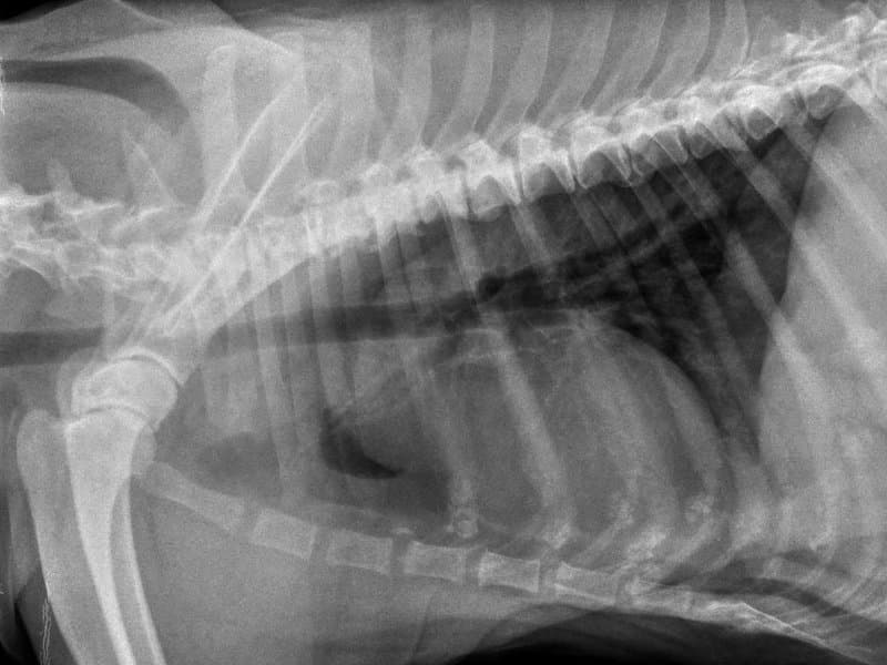
DX · lateral
✓ Peer-reviewed
Canine Thorax
Canine thoracic radiograph in right-lateral recumbency. Air-filled lungs read as low-density (dark) against the soft-tissue silhouette of heart, great vessels and diaphragm.
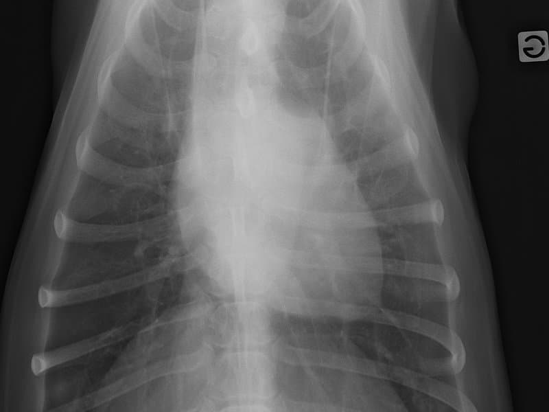
DX · VD
✓ Community
Canine Thorax
Canine thoracic radiograph in ventrodorsal projection. Lung fields are symmetric left and right of the cardiac silhouette; sternum overlies the spine.
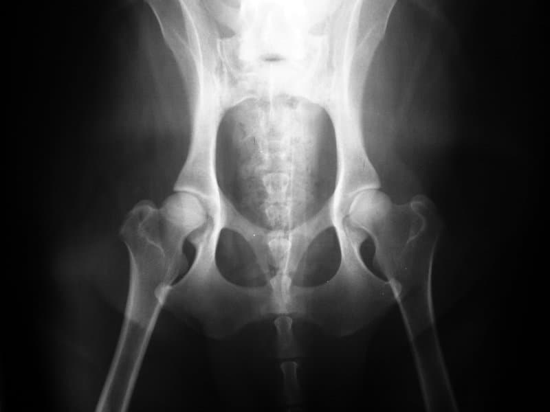
DX · VD
✓ Community
Canine Pelvis
Canine pelvic radiograph in extended-leg ventrodorsal projection — the standard view for hip joint evaluation. Symmetric placement of femoral heads in acetabula is the baseline for Norberg-angle measurement.
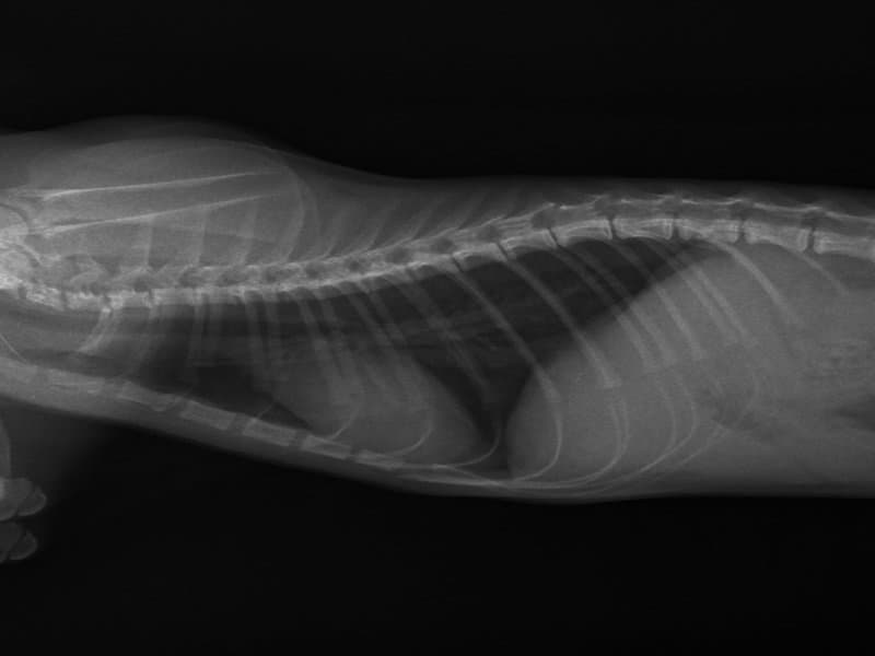
DX · lateral
✓ Peer-reviewed
Feline Thorax
Feline thoracic radiograph in right-lateral recumbency. Compared with canine the cardiac silhouette is more elongated, vertebral heart score reference range differs (feline ~6.7–8.1 vs canine ~8.5–10.5).
DX · lateral
✓ Community
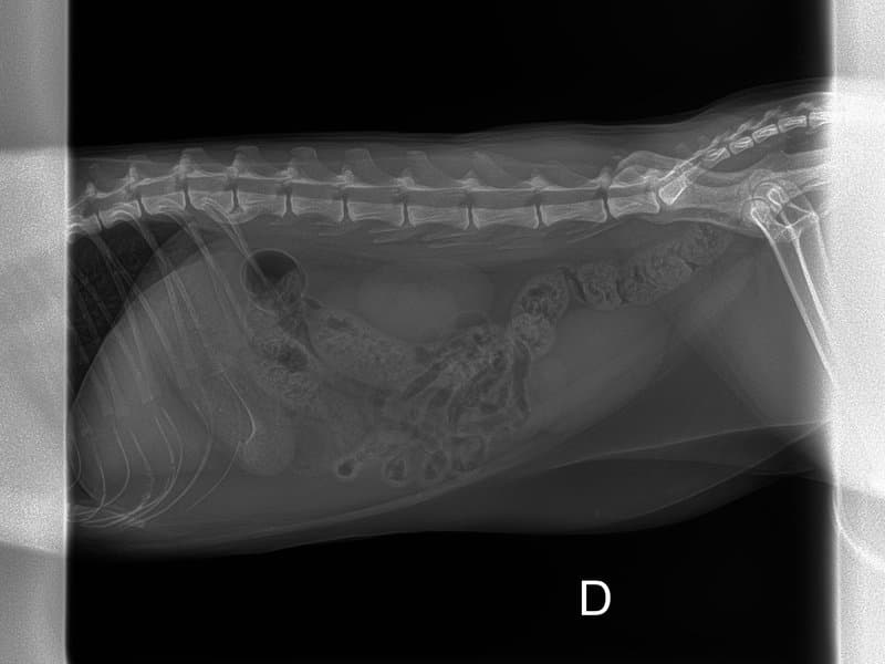
Feline Abdomen
Feline abdominal radiograph in right-lateral recumbency. Smaller frame than canine; renal silhouettes are typically more conspicuous due to retroperitoneal fat.
DX · VD
✓ CUVET
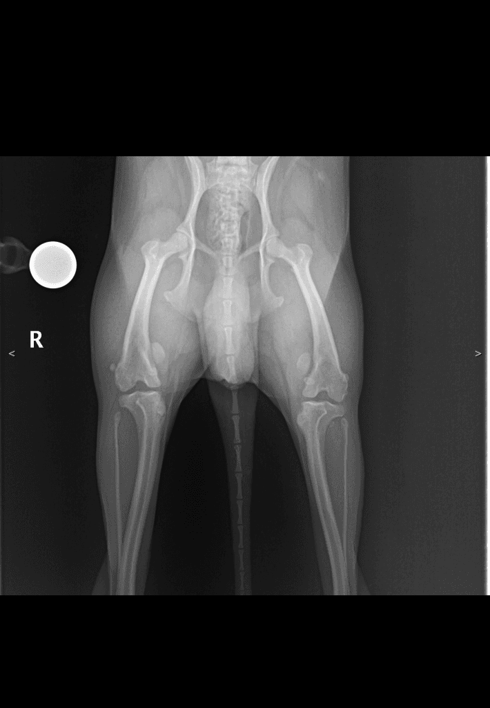
Canine Pelvis
Canine extended-leg ventrodorsal pelvis from the CUVET teaching set. Symmetric femoral-head placement in acetabula — Norberg-angle baseline view. Right-side anatomy marker (R) visible.
DX · DV
✓ CUVET
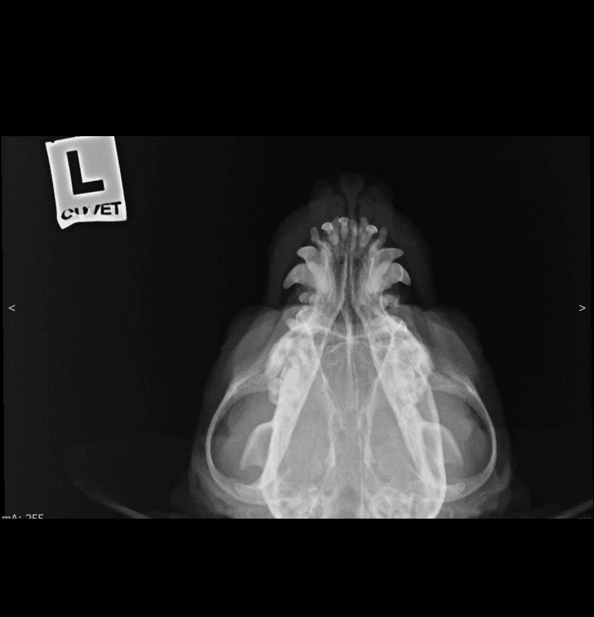
Feline Skull
Feline dorsoventral skull from the CUVET teaching set. Symmetric tympanic bullae, dental arcades and zygomatic arches in plain view. Lead L-marker visible top-left.
DX · lateral
✓ CUVET
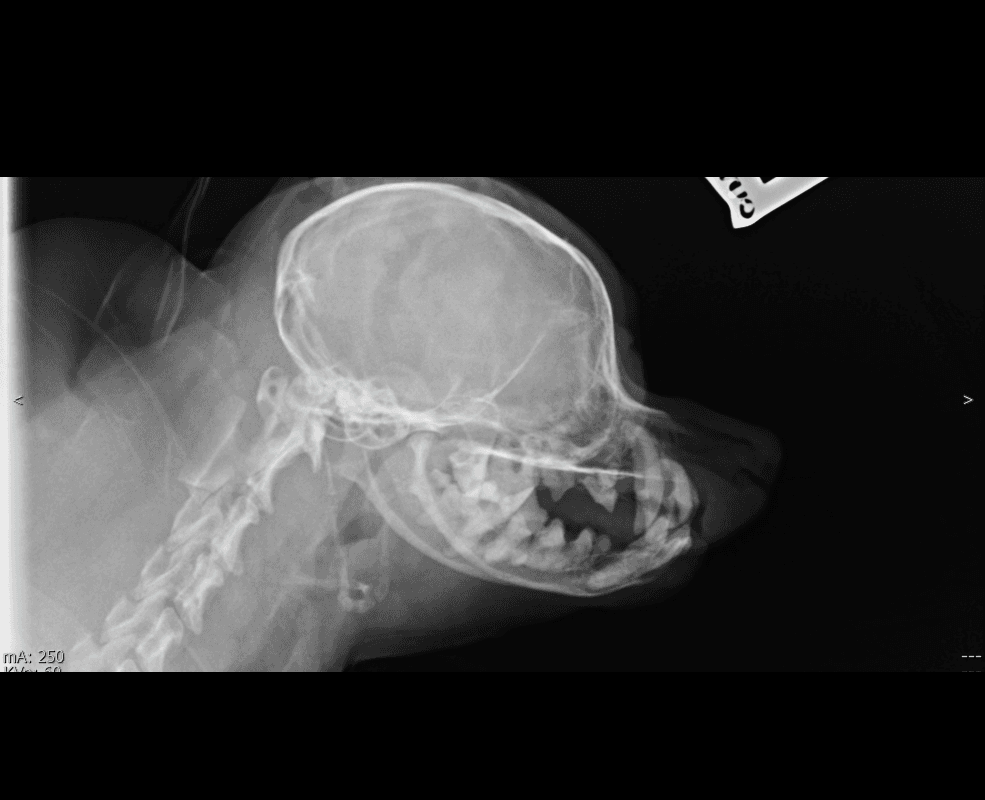
Feline Skull
Feline lateral skull from the CUVET teaching set — rounded cranial vault and short rostrum typical of feline conformation. Useful contrast against the longer-muzzled canine lateral for skull-shape comparison. (Breed unknown; cephalic conformation not asserted from a single anonymized view.)
DX · lateral
✓ CUVET
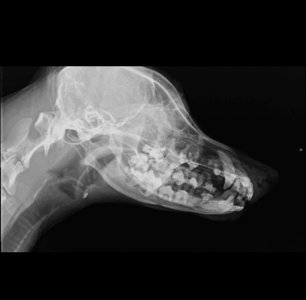
Canine Skull
Canine lateral skull from the CUVET teaching set — elongated muzzle with complete adult dental arcades (longer-faced conformation). Breed unknown; specific cephalic class not asserted from a single anonymized view.
DX · lateral
✓ CUVET
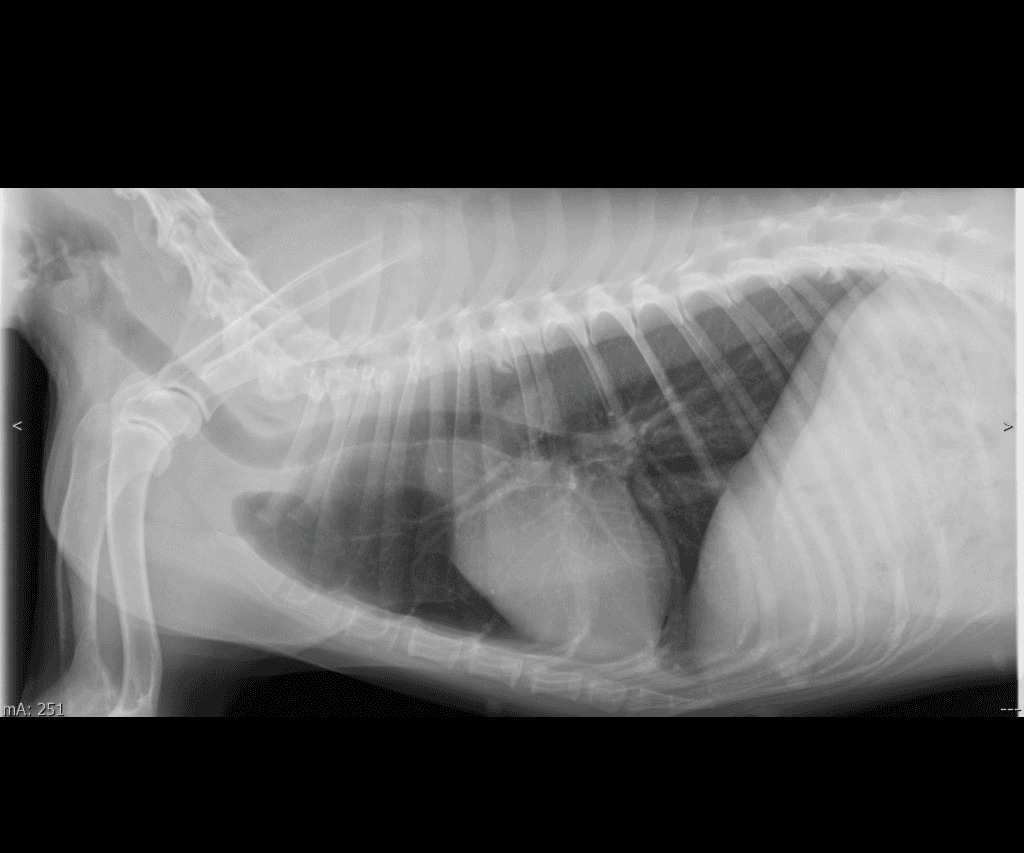
Canine Thorax
Canine lateral thorax from the CUVET teaching set. Cardiac silhouette, tracheal column, and diaphragmatic line in profile — VHS-compatible view for vertebral heart score practice (canine reference range 8.5–10.5).
DX · lateral
✓ CUVET
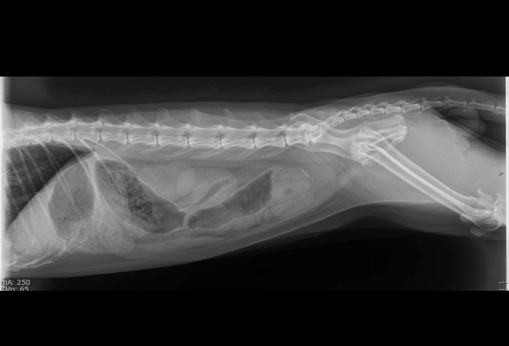
Canine Abdomen
Canine lateral abdomen from the CUVET teaching set. Vertebral column dorsally, gas-filled bowel loops + serosal silhouettes ventrally, pelvic inlet caudally. The reference layout for evaluating gas pattern, organ silhouette, and serosal detail.
DX · VD
✓ CUVET
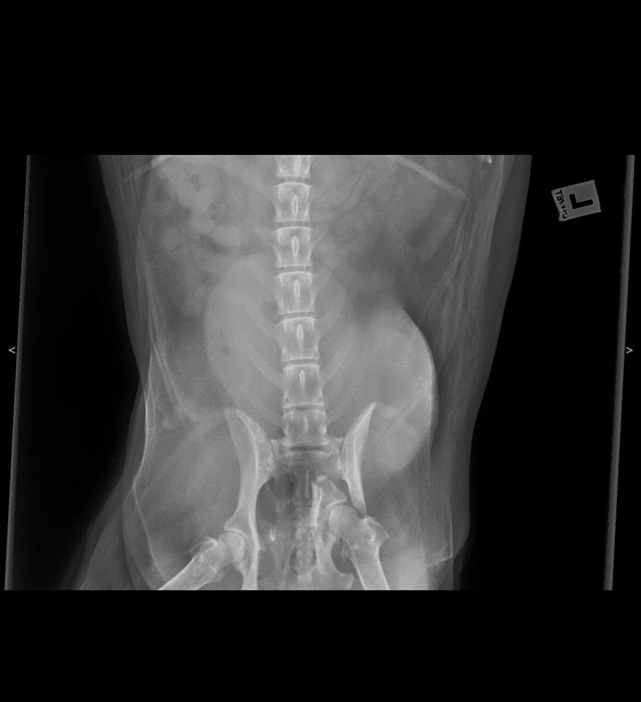
Canine Abdomen
Canine ventrodorsal abdomen from the CUVET teaching set. Vertebrae down the midline, symmetric kidneys flanking, intestinal gas patterns, and sacroiliac + femoral heads at the pelvic inlet. Pair with the lateral view for two-plane abdominal evaluation.
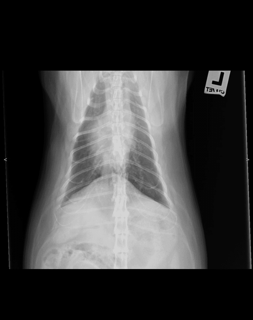
DX · VD
✓ CUVET
Canine Spine
Canine VD spanning the thoracic vertebral column into the cranial lumbar (frame ends mid-lumbar), ribs symmetric on both sides — useful for screening vertebral alignment, intervertebral disc-space uniformity, and vertebral body shape across the thoracolumbar junction.
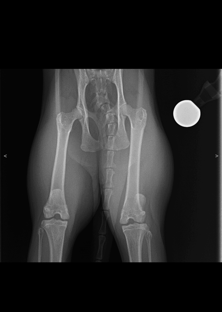
DX · VD
✓ CUVET
Canine Hindlimb
Canine ventrodorsal pelvis + bilateral stifles from the CUVET teaching set. Extends a standard Norberg-positioning VD caudally to capture both femorotibial joints — femoral condyles, tibial plateaus, and fabellae visible on both sides. Useful for stifle symmetry screening and as a one-image hindlimb reference.
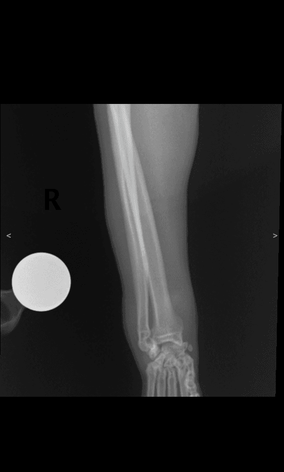
DX · lateral
✓ CUVET
Canine Forelimb
Canine lateral antebrachium (radius + ulna) and carpus from the CUVET teaching set. Full forearm shaft visible with the carpal joints at the distal end. Reference for forelimb-shaft alignment and carpal-row anatomy.
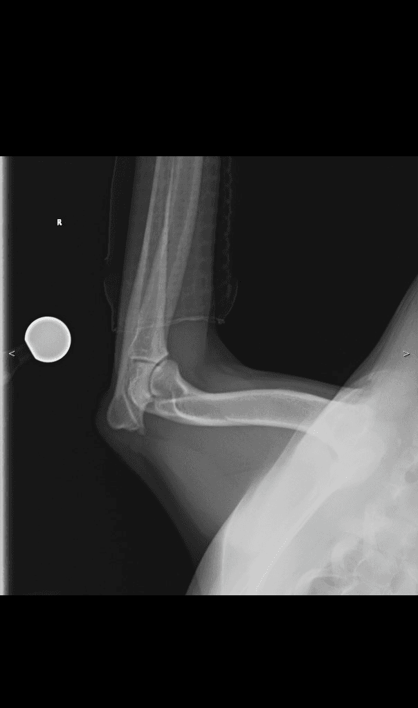
DX · lateral
✓ CUVET
Canine Forelimb
Canine lateral elbow (humero-radio-ulnar joint) from the CUVET teaching set. Joint flexed at ~110° in this projection, with the humeral condyle proximally, anconeal process projecting behind the joint, and radius + ulna distally. Body-wall shadow visible caudoventrally (right side of image).
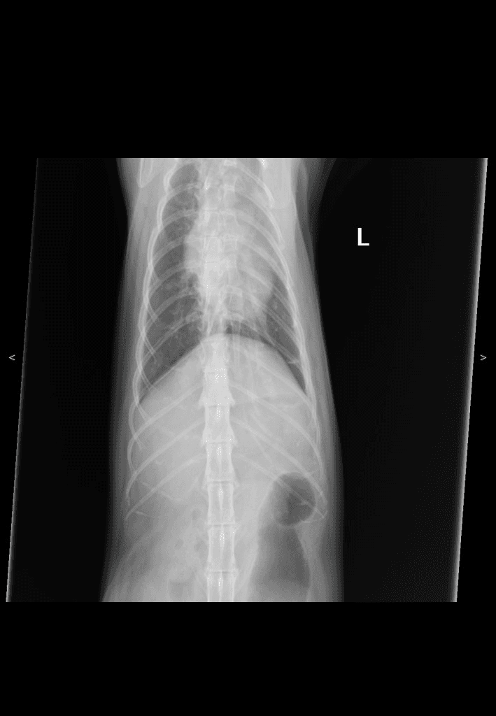
DX · VD
✓ CUVET
Canine Thorax
Canine ventrodorsal thorax from the CUVET teaching set. Symmetric ribs left + right of midline, cardiac silhouette centred, spine running down midline. Pair with cuvet-canine-thorax-lat-001 for two-plane thoracic evaluation.
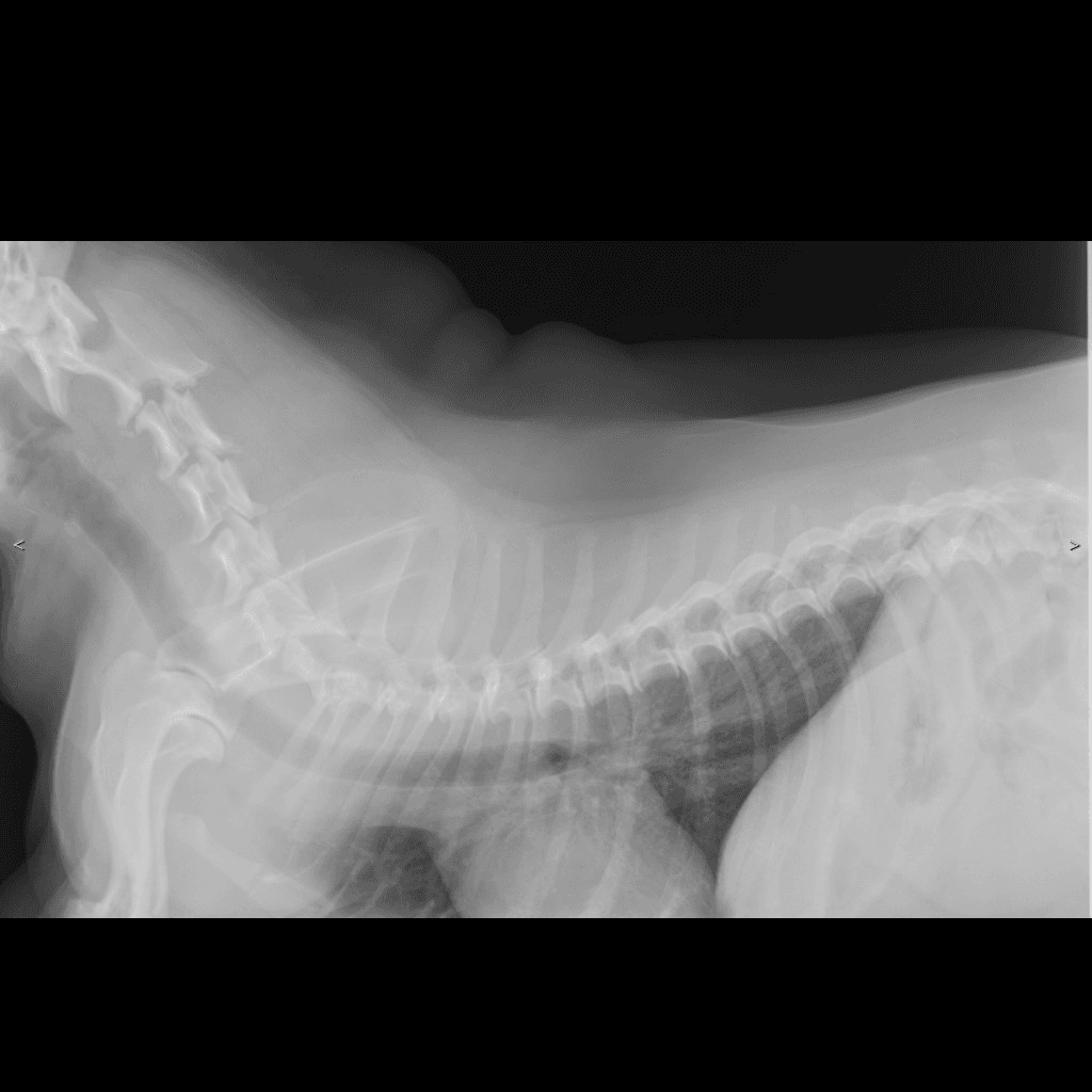
DX · lateral
✓ CUVET
Canine Spine
Canine cervicothoracic lateral from the CUVET teaching set (study labeled 'Chest'). The cervical vertebral column C1–C7 is clearly visible curving from the skull base through the cervicothoracic junction into the cranial thorax — a reference for counting cervical vertebrae and reading the normal cervical curvature. Anonymized via the CUVET 4-pass scrubber pipeline.
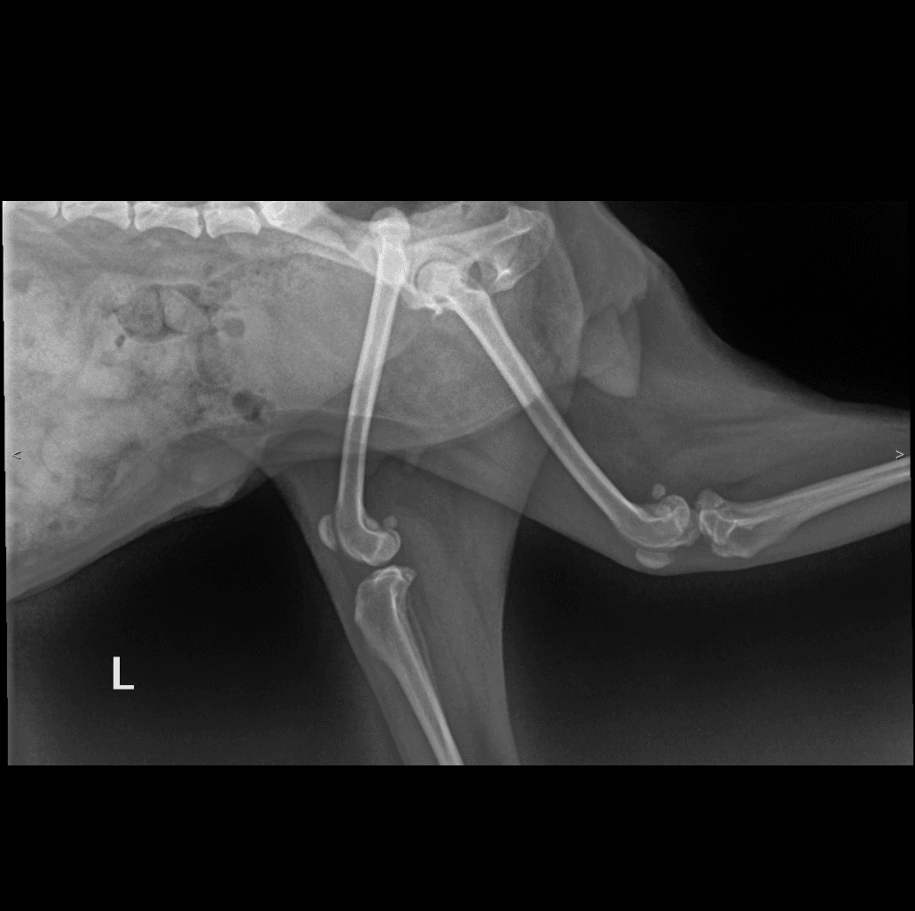
DX · lateral
✓ CUVET
Canine Pelvis
Canine lateral pelvis / pelvic limb from the CUVET teaching set (study labeled 'Hip-joint'). Hip joint, femur, stifle, and proximal tibia are visible in one lateral projection — the first lateral pelvis in the atlas to pair with the VD pelvis views, and the first lateral look at the stifle. Anonymized via the CUVET 4-pass scrubber pipeline.
19 / 19 atlas tiles are real diagnostic radiographs (no AI fill). Sources: peer-reviewed VetXRay (Zenodo, CC BY 4.0), Wikimedia Commons (CC BY / CC BY-SA), and anonymized CUVET teaching cases (Aj.-approved · PII-scrubbed via 4-pass pipeline).
Coverage gaps we openly track: a confirmed-normal stifle (lateral) — an isolated stifle lateral is now live as a post-operative case under Cases, but a normal-anatomy reference is still pending. Trust the badge, not the filename. ดูแหล่งข้อมูลทั้งหมด →