


Canine Abdomen — lateral
DX · digital X-ray
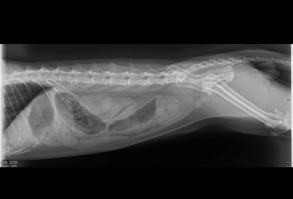
DX · Canine · Abdomen · lateral✓ cuvet-internal reference
01 /What's visible
Canine lateral abdomen from the CUVET teaching set. Vertebral column dorsally, gas-filled bowel loops + serosal silhouettes ventrally, pelvic inlet caudally. The reference layout for evaluating gas pattern, organ silhouette, and serosal detail.
02 / Landmarks
- Vertebral column (T13–L7)
- Diaphragm (cranial border)
- Liver silhouette (caudal to diaphragm)
- Gastric gas
- Small intestinal loops
- Colon
- Urinary bladder
- Pelvic inlet (caudal limit)
03 /Source & license
- License
- Educational use, CUVET-internal
- Credibility
- cuvet-internal
- Attribution
- Courtesy of Small Animal Hospital of Chulalongkorn University · DI Unit · anonymized for veterinary education with permission
04 / Next — apply to a case
Open in DICOM viewer — Canine lateral abdomen (CUVET)paired case
Same anonymized radiograph wrapped as DICOM — practice Norberg / VHS / Length measurements live on this image.
Clinical case library — Abdomenread abnormal next
Now that you have a baseline for normal abdomen, open a case from the library to spot what's different.
05 / Other abdomen views
DX · lateral
✓ Community
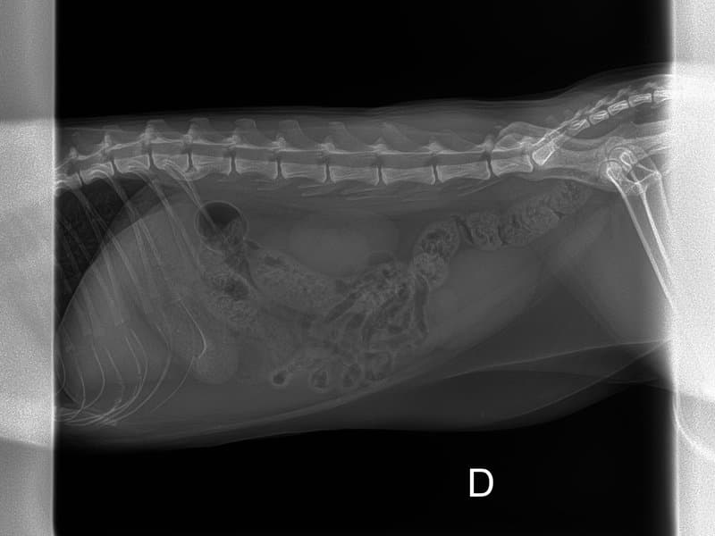
Feline Abdomen
Feline abdominal radiograph in right-lateral recumbency. Smaller frame than canine; renal silhouettes are typically more conspicuous due to retroperitoneal fat.
DX · VD
✓ CUVET
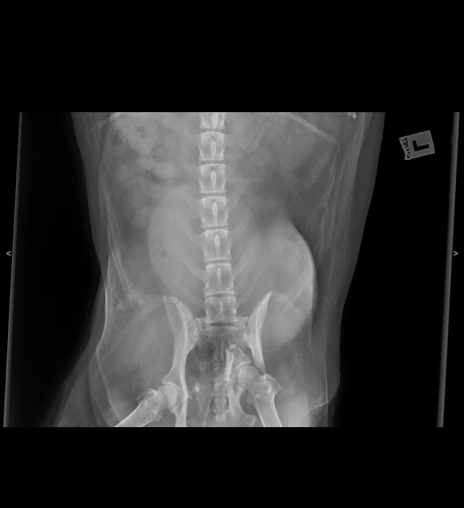
Canine Abdomen
Canine ventrodorsal abdomen from the CUVET teaching set. Vertebrae down the midline, symmetric kidneys flanking, intestinal gas patterns, and sacroiliac + femoral heads at the pelvic inlet. Pair with the lateral view for two-plane abdominal evaluation.