


Canine Thorax — lateral
DX · digital X-ray
DX · Canine · Thorax · lateral✓ Peer-reviewed reference
01 /What's visible
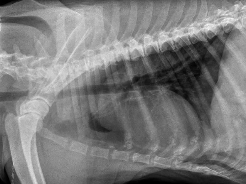
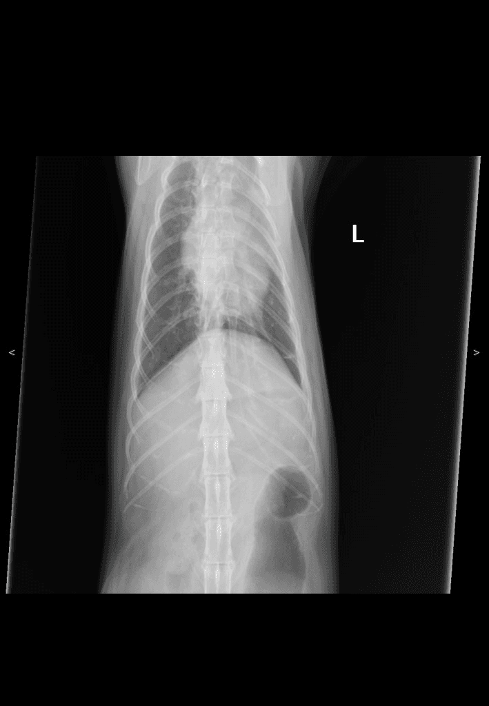
Canine thoracic radiograph in right-lateral recumbency. Air-filled lungs read as low-density (dark) against the soft-tissue silhouette of heart, great vessels and diaphragm.
02 / Landmarks
- Cardiac silhouette
- Diaphragmatic line
- Tracheal column
- Caudal vena cava
- Vertebral column dorsally
03 /Source & license
- License
- CC BY 4.0
- Credibility
- peer-reviewed
- Attribution
- VetXRay — 9,882 manually annotated canine and feline thoracic radiographs · Zenodo · DOI:10.5281/zenodo.19051776 · CC BY 4.0
- Source
- https://zenodo.org/records/19051776 ↗
04 / Next — apply to a case
Clinical case library — Thoraxread abnormal next
Now that you have a baseline for normal thorax, open a case from the library to spot what's different.
05 / Other thorax views
DX · VD
✓ Community
Canine Thorax
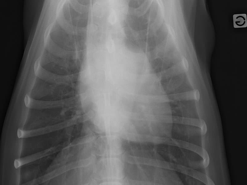
Canine thoracic radiograph in ventrodorsal projection. Lung fields are symmetric left and right of the cardiac silhouette; sternum overlies the spine.
DX · lateral
✓ Peer-reviewed
Feline Thorax
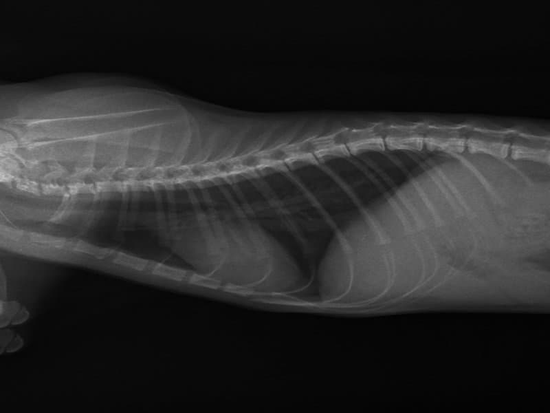
Feline thoracic radiograph in right-lateral recumbency. Compared with canine the cardiac silhouette is more elongated, vertebral heart score reference range differs (feline ~6.7–8.1 vs canine ~8.5–10.5).
DX · lateral
✓ CUVET
Canine Thorax
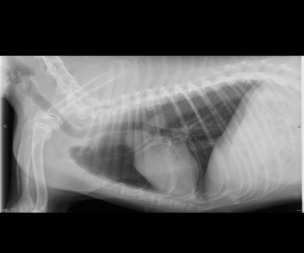
Canine lateral thorax from the CUVET teaching set. Cardiac silhouette, tracheal column, and diaphragmatic line in profile — VHS-compatible view for vertebral heart score practice (canine reference range 8.5–10.5).
DX · VD
✓ CUVET
Canine Thorax
Canine ventrodorsal thorax from the CUVET teaching set. Symmetric ribs left + right of midline, cardiac silhouette centred, spine running down midline. Pair with cuvet-canine-thorax-lat-001 for two-plane thoracic evaluation.