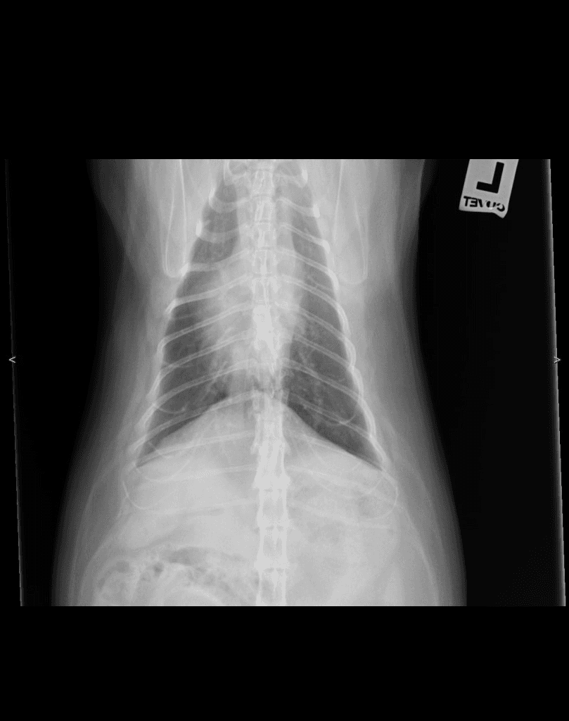
Canine Spine — lateral
DX · digital X-ray
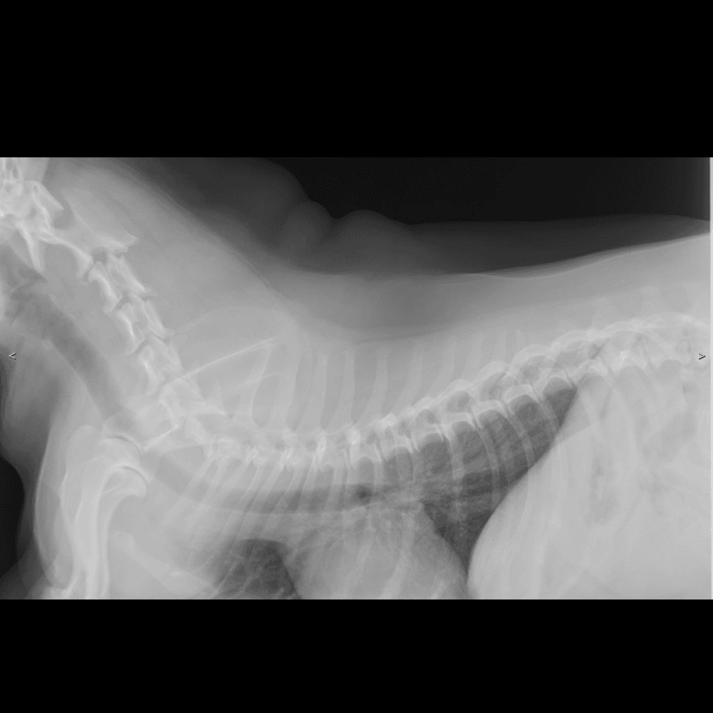
DX · Canine · Spine · lateral✓ cuvet-internal reference
01 /What's visible
Canine cervicothoracic lateral from the CUVET teaching set (study labeled 'Chest'). The cervical vertebral column C1–C7 is clearly visible curving from the skull base through the cervicothoracic junction into the cranial thorax — a reference for counting cervical vertebrae and reading the normal cervical curvature. Anonymized via the CUVET 4-pass scrubber pipeline.
02 / Landmarks
- Cervical vertebrae C1 (atlas) → C7
- Cervicothoracic junction (C7–T1)
- Cranial thoracic vertebrae + ribs
- Trachea (lucent column ventral to the spine)
- Cardiac silhouette (cranial thorax)
- < / > positioning markers (lateral edges)
03 /Source & license
- License
- Educational use, CUVET-internal
- Credibility
- cuvet-internal
- Attribution
- Courtesy of Small Animal Hospital of Chulalongkorn University · DI Unit · anonymized for veterinary education with permission
04 / Next — apply to a case
Open in DICOM viewer — Canine cervical spine lateral (CUVET)paired case
Same anonymized radiograph wrapped as DICOM — practice Norberg / VHS / Length measurements live on this image.
Clinical case library — Spineread abnormal next
Now that you have a baseline for normal spine, open a case from the library to spot what's different.