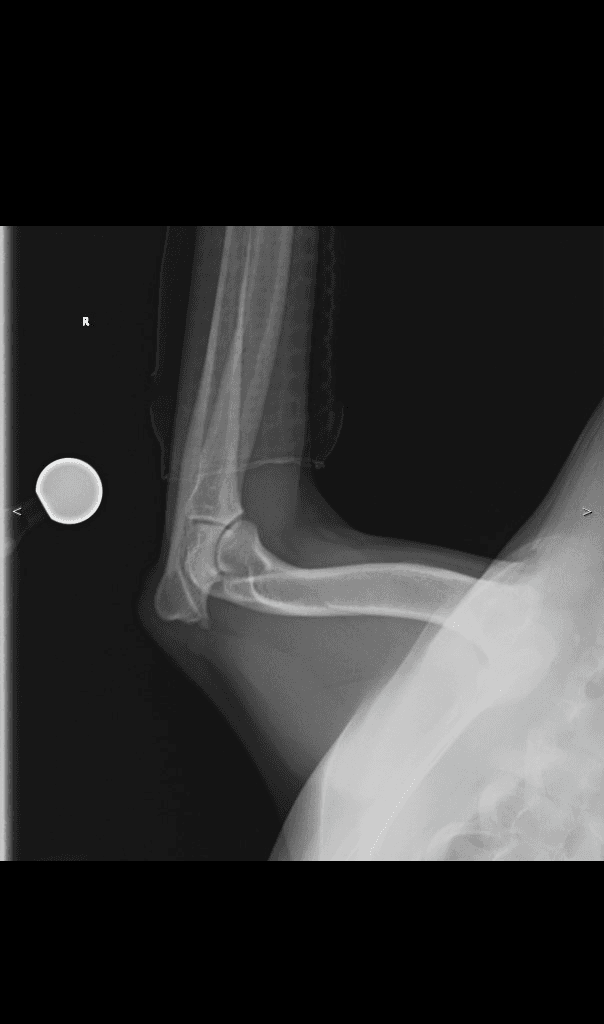
Canine Forelimb — lateral
DX · digital X-ray
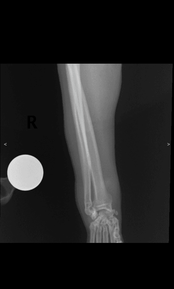
DX · Canine · Forelimb · lateral✓ cuvet-internal reference
01 /What's visible
Canine lateral antebrachium (radius + ulna) and carpus from the CUVET teaching set. Full forearm shaft visible with the carpal joints at the distal end. Reference for forelimb-shaft alignment and carpal-row anatomy.
02 / Landmarks
- Radius (cranial bone, slimmer)
- Ulna (caudal bone, with olecranon proximally)
- Antebrachiocarpal joint (distal radius)
- Carpal bones (proximal + distal rows)
- Metacarpal bases
- R anatomy marker (top-left)
03 /Source & license
- License
- Educational use, CUVET-internal
- Credibility
- cuvet-internal
- Attribution
- Courtesy of Small Animal Hospital of Chulalongkorn University · DI Unit · anonymized for veterinary education with permission
04 / Next — apply to a case
Open in DICOM viewer — Canine forelimb lateral · antebrachium + carpus (CUVET)paired case
Same anonymized radiograph wrapped as DICOM — practice Norberg / VHS / Length measurements live on this image.
Clinical case library — Forelimbread abnormal next
Now that you have a baseline for normal forelimb, open a case from the library to spot what's different.