


Feline Skull — DV
DX · digital X-ray
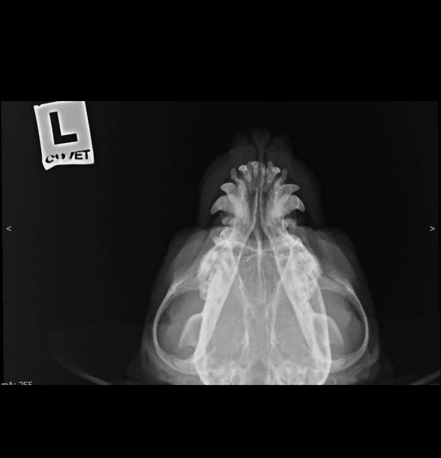
DX · Feline · Skull · DV✓ cuvet-internal reference
01 /What's visible
Feline dorsoventral skull from the CUVET teaching set. Symmetric tympanic bullae, dental arcades and zygomatic arches in plain view. Lead L-marker visible top-left.
02 / Landmarks
- Tympanic bullae (paired, symmetric)
- Zygomatic arches
- Maxillary + mandibular dental arcades
- Nasal cavity midline
- Cranial vault outline
- Lead L marker (CUVET institutional, top-left)
03 /Source & license
- License
- Educational use, CUVET-internal
- Credibility
- cuvet-internal
- Attribution
- Courtesy of Small Animal Hospital of Chulalongkorn University · DI Unit · anonymized for veterinary education with permission
04 / Next — apply to a case
Open in DICOM viewer — Feline skull DV (CUVET) · first feline DICOM casepaired case
Same anonymized radiograph wrapped as DICOM — practice Norberg / VHS / Length measurements live on this image.
Clinical case library — Skullread abnormal next
Now that you have a baseline for normal skull, open a case from the library to spot what's different.
05 / Other skull views
DX · lateral
✓ CUVET
Feline Skull
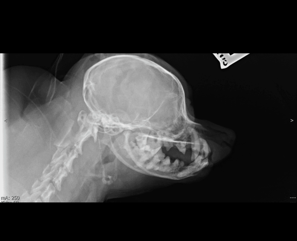
Feline lateral skull from the CUVET teaching set — rounded cranial vault and short rostrum typical of feline conformation. Useful contrast against the longer-muzzled canine lateral for skull-shape comparison. (Breed unknown; cephalic conformation not asserted from a single anonymized view.)
DX · lateral
✓ CUVET
Canine Skull
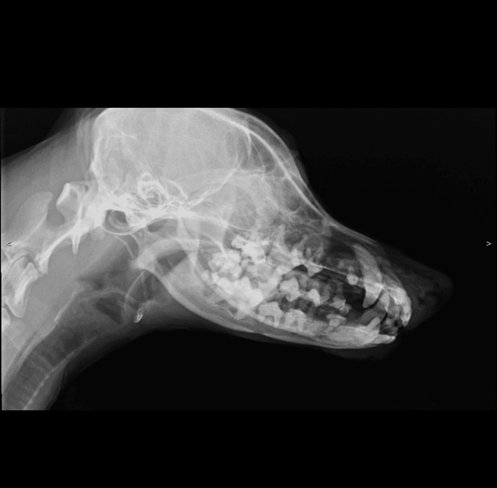
Canine lateral skull from the CUVET teaching set — elongated muzzle with complete adult dental arcades (longer-faced conformation). Breed unknown; specific cephalic class not asserted from a single anonymized view.